From now on, you can also visit us in our online consultation. Using a camera and microphone, we can make an initial assessment of your wishes and symptoms and agree on subsequent steps. Just visit us at https://arzt-direkt.com/praxis-gussmann-drecoll. We look forward to seeing you. Your team at the dermatology practice B18 Gussmann+Drecoll
Read MoreOur consultation hours
| Monday | 9:00 – 13:0015:00 – 18:00 |
|---|---|
| Tuesday | 9:00 – 13:0015:00 – 18:00 |
| Wednesday | 9:00 – 13:00 |
| Thursday | 9:00 – 13:0015:00 – 18:00 |
| Friday | 9:00 – 13:00 |
How to reach us
Dermatologische Praxis B18
Gussmann & Eydam
Badensche Strasse 18
10715 Berlin-Wilmersdorf
Telefon: 030 814 869 30
Fax: 030 814 869 31
E-Mail: info@dermatologen-b18.de
Transport connection
U-Bahn: U7, U9 to U-Station Berliner Straße
Approach: Gesundheitszentrum B18, Badensche Straße 18, Berlin-Wilmersdorf
It goes without saying that our house is handicapped accessible.
Dermatology practice B18
Medical treatments
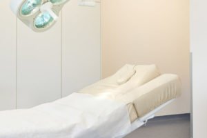
Both doctors perform all operations themselves in the practice’s modern operating theatre. They place particular emphasis on gentle operations and an aesthetic result.
Since all operations are performed under local anaesthesia, every patient can leave the practice after the operation.
Both doctors have a high level of expertise after having worked in a leading position at the Charité Hospital in Berlin.
One of the most modern and safest methods in skin cancer screening is computer-assisted reflected-light microscopy, which is used for the early detection of conspicuous skin changes and image documentation. With the help of this procedure, skin changes can be examined, assessed and, above all, documented in the long term much better.
With digital reflected-light microscopy, the patient can follow the entire examination on a computer screen. The conspicuous skin changes are recorded with a special high-performance camera with up to 70-fold magnification and optimally displayed on the screen.
The computer evaluates the image data based on certain factors (e.g. extent of the skin change, shape, pigmentation, etc.) and determines a risk score. This gives an initial assessment of whether the skin change can be classified as harmless, suspicious or dangerous.
The image data of conspicuous skin changes can also be saved and compared with the digital images of the last examination at the next check-up. In this way, even the slightest changes can be detected that are not visible to the naked eye.
Thanks to precise diagnostics and the best possible control, unnecessary biopsies and/or operations can be avoided in many cases with the help of computer-assisted reflected-light microscopy.
In case of doubt, however, only a biopsy (tissue sample) can provide definitive clarity as to whether a conspicuous skin change is benign or malignant. Despite increased diagnostic certainty, computer-assisted image analysis cannot replace the knowledge and experience of a qualified dermatologist in the assessment of moles, but it can support him in his work as best as possible.
If a mole has certain criteria that make it difficult to draw a clear line to melanoma, for example in the context of hair cancer screening, we can use Nevisense from Scibase for diagnostics. In cases of doubt, this device provides us with data from deeper layers of the pigment spot within a few minutes and thus helps to avoid unnecessary operations.
Further information under: https://scibase.com/clinical-benefits/
With OCT (Optical Coherence Tomography) we have a procedure at our disposal with which, similar to an ultrasound device, vertical sectional images of the skin can be created.
This enables us to diagnose skin tumours, such as basal cell or squamous cell carcinoma and numerous benign lesions, quickly and painlessly. With a penetration depth of about 1.5 mm, numerous superficial structures and processes of the skin can be examined.
We use this procedure for diagnostics, expansion diagnostics before planned operations and for therapy control after treatments have been carried out.
Photodynamic therapy (PDT) is a modern and proven procedure for the treatment of ‘light skin cancer’ and especially its precursors (so-called actinic keratoses). The great advantage lies in the possibility of effectively treating larger areas, such as the scalp or the entire face or décolleté.
As the penetration depth into the skin is limited to a few millimetres, the treatment is also reserved exclusively for superficial skin tumours and their precursors.
In the first step, the treatment area is treated with a cream, which is left on for approx. 3 hours. You do not need to be present in our practice during this time.
In the second step, the area is exposed to a cold, red light. During this exposure phase, the skin feels warm and the skin cancer cells are damaged. Healthy skin cells are spared with this treatment method. In the case of extensive findings, the treatment can also be painful. But even here we have ways of making the treatment bearable.
During the entire treatment time, which is usually between 10 and 60 minutes, you will be carefully looked after by our qualified staff.
The damaged skin cancer cells are then shed over the next few days. During this process, which is completed after a few days (usually after approx. 1 week), individual redness and crusts form.
Classic PDT using a PDT lamp can be carried out all year round.
ADL PDT (Artificial Daylight PDT) can be carried out at any time of the year in a controlled manner. Large treatment areas can be covered in one session. Additional cooling is not necessary. This is possible both in our practice and as DL-PDT (Daylight PDT) outdoors in the summer months.
The virtually painless treatment is highly effective.
As with conventional lamp therapy, a medication is first applied to the areas to be treated, which then unfolds its effect together with the light.
Following the treatment, depending on the individual light damage, a mild to moderate, rarely also pronounced inflammatory reaction with severe reddening and even crust formation develops.
The more pronounced the existing photodamage, the stronger these skin symptoms are and they usually disappear completely within 7-14 days.
Modern lasers have become an indispensable part of dermatology. They are used in both the medical and aesthetic fields.
In the context of medical applications, the correct diagnosis must first be made by the specialist. Then lasers can be a useful and elegant treatment method to remove superficial skin lesions.
Lasers can also be an alternative to conventional surgery.
Excessive sweating (hyperhidrosis) can occur on different parts of the body and can be very annoying, even downright interfering with everyday life and social life. Common locations are the armpits, hands and feet.
There are a number of ways to achieve significant relief. We will be happy to advise you individually on various treatment methods.
Varicose veins are often not only cosmetically disturbing, but can also pose a health risk. An initial examination is carried out in the practice by means of a Doppler examination.
Acne is a disease of the skin glands and hair follicles. Comedones (blackheads) are also found. Most people develop varying degrees of acne during puberty.
For some, only small inflammatory papules and pustules appear, especially on the face, décolleté and back. Others develop extensive, large inflammatory lumps, sometimes with abscess formation.
The disease is often accompanied by considerable suffering both during the acute phase of the disease and after healing. Acne can also recur in adulthood.
The aim is therefore a multi-phase treatment appropriate to the stage, which either primarily heals acute inflammation or reduces secondary scars and pigmentation disorders.
Micro-Botox on the face is also particularly suitable for reducing sebum production in order to achieve a reduction in acne.
We offer you a range of different, individualised treatment options, also in combination with our medical cosmetics.
Rosacea is a skin disease that usually occurs on the midface and often appears similar to acne. It progresses in different stages and the clinical appearance ranges from slight redness, increased vascularisation (‘couperose’) to severely inflamed papules and pustules or the formation of a rhinophyma (so-called bulbous nose). The eyes can also be affected, in which case ophthalmological treatment is sometimes necessary.
Treatment depends on the stage and ranges from topical treatment to systemic treatment with antibiotics.
Dilated vessels (so-called couperose) can be treated very well with the KTP laser. The result is visible immediately after the treatment.
Rhinophyma can be treated effectively with the CO2 laser. Depending on the degree of severity, a local anaesthetic is usually sufficient.
By using different lasers and our medical cosmetics, we can also realise individual treatment plans to improve the appearance of your skin in the long term.
Micro-Botox can also be a helpful treatment for rosacea. This is a new, innovative therapeutic approach. The typical redness can be reduced.
In our practice, we offer comprehensive solutions for your foot health. Together with our medical foot care, our services include the following:
- Nail fungus – diagnosis and treatment to restore healthy nails.
- Nail growth disorders – professional treatment to alleviate symptoms.
- Ingrown toenails – customised solutions, including the application of nail braces to eliminate ingrowth. These are glued to the nail and grow out with the nail.
Our aim is to provide you with professional and personalised care to promote your foot health.
At the B18 dermatological practice, we offer you comprehensive medical cosmetic solutions that are tailored to your individual skin needs. Our services include:
- Acne treatments: – We develop customised therapy concepts to effectively combat acne and its consequences to help you achieve a clear complexion.
- Rosacea therapy: Our treatments specifically designed for rosacea help to alleviate the symptoms and improve the appearance of the skin.
- Professional facial care: For patients* who value qualified skin care, we offer individualised treatments that serve to both prevent and regenerate your skin.
At the B18 dermatological practice, your needs take centre stage. Make an appointment to receive a personalised consultation and find the right treatment for you.
Aesthetic procedures
The areas of application of the fractional CO2 laser (Fraxel laser) are manifold.
It is used to improve the skin structure (reduction of small wrinkles and tightening of the skin through the formation of collagen, among other things), tightening of the lower eyelids and gives the skin a more even complexion by removing superficial pigment spots and irregularities.
Acne and chicken pox scars, small wrinkles, especially in the area of the lower eyelids or skin rejuvenation of the face, neck, décolleté or hands are among the classic areas of application with usually impressive results. Depending on the indication, one or more treatments are required to achieve the desired result.
After a short healing phase (usually a few days), the first treatment results are already visible.
All aesthetic treatments are preceded by a detailed consultation and information session. You will then receive a written information sheet, an individual cost plan and a treatment contract.
The KTP laser has a wavelength of 532 nm and is therefore very well suited for the treatment of superficially dilated vessels and haemangiomas, which appear as so-called “couperose” on the face.
Small warts, fibromas and other benign, disturbing skin changes can also be gently removed with the KTP laser.
The short light pulse penetrates the upper layer of the skin so that the energy is first absorbed by the red blood pigment in the blood vessels.
The vessels are heated, contract, no longer receive blood and disappear.
This effect is already visible directly after the treatment. Reddening in the treated area immediately after the treatment is completely normal and usually disappears completely after a few hours.
Occasionally, however, small crusts may appear that fall off on their own after a few days. Depending on the type and extent of vascular dilatation, several treatments may be necessary.
All aesthetic treatments are preceded by a detailed consultation and information session. You will then receive a written information sheet, an individual cost plan and a treatment contract.
The Q-switched ruby laser for pigmented skin lesions and multi-coloured tattoos has an optimal wavelength of 694 nm and extremely short pulses of only 20 ns. This makes it one of the safest and most precise treatment solutions for benign pigmented skin lesions and for removing all types of tattoos, whether decorative or dirty, professional or amateur, or permanent make-up.
Whether in preparation for a cover up or for permanent tattoo removal, the ruby laser is ideally suited. As a rule, the treatment sessions are carried out at intervals of at least 6 weeks. The interval between laser and cover-up should be at least 4 months.
Disturbing benign pigment spots (e.g. “age spots”) can be removed gently and permanently. The pigment is broken down into its smallest pieces and removed by the body’s own macrophages. This explains the breaks in treatment that are always necessary.
All aesthetic treatments are preceded by a detailed consultation and explanatory talk. You will then receive a written information sheet, an individual cost plan and a treatment contract.
Mimic muscles are the muscles of the face that intentionally or unintentionally bring movement to our face.
Over the years, very strong and active muscles can lead to the formation of wrinkles in the overlying skin, which may give us an expression that outwardly reflects our basic mood incorrectly. This can make us appear unintentionally angry, sad or stressed.
Small wrinkles around the eyes caused by frequent, unconscious squinting may also be distracting.
Botulinum toxin enables us to give you back the relaxed facial expression you desire. Depending on the dose, the temporary interruption of the signalling pathway between nerve and muscle leads to a reduction in muscle activity and consequently to a relaxation of the overlying skin.
The wrinkles are reduced or even disappear completely. As a rule, the treatment is repeated every 3-6 months to maintain the effect.
In addition to the classic indications such as frown lines, forehead wrinkles and wrinkles around the eyes, we also treat other localisations.
These include wrinkles in the neck area, reduction of undesirable dimpling on the chin due to pronounced muscular activity (cobblestone chin), bunny lines in the nose area, ‘lip flip’ of the upper lip, micro-botox on the face,
In addition to gentle smoothing, this is also particularly suitable for reducing sebum production and thus improving acne symptoms. Treatment with botulinim toxin is also an innovative treatment method for reducing the redness of rosacea.
In addition to these indications relating to the skin, there are other established treatment options in other areas.
These include, for example, the treatment of the masseter muscle to reduce teeth grinding (bruxism) and thus contribute to dental health.
Botulinum toxin can also be used to reduce the sometimes disturbing hypertrophy (enlargement) of the masseter muscle, which is caused by increased muscle tone and visually widens the face (facial slimming)
All aesthetic treatments are preceded by a detailed consultation and information session. You will then receive a written information sheet, a personalised cost plan and a treatment contract.
Hyaluronic acid (filler)
Multiple indications:
- Sagging cheeks
- Hollow cheeks
- sagging neckline
- droopy lower eyelids
- dark tear trough
- sagging facial skin
Advantages of thread lifting:
- Less stress than a traditional facelift
- Fast healing
- Short treatment time (only about 15 to 45 minutes)
- Low risk of pain and inflammation
The treatment area is locally anaesthetised and the absorbable thread is inserted into the tissue. The type and number of sutures to be used are determined in advance in a detailed consultation and information session.
This minimally invasive procedure builds new connective tissue around the thread, making the skin firmer and smoother. Certain types of thread can also lift skin and subcutaneous tissue, returning it to approximately its original position.
This new and gentle method usually leaves no wounds or scars. The threads are absorbed after a certain time (depending on the type of thread), i.e. they dissolve. The results are visible for months to years, depending on the thread and indication.
All aesthetic treatments are preceded by a detailed consultation and information session. You will then receive a written information sheet, an individual cost plan and a treatment contract.
A ‘vampire lift’ or PrP (platelet-rich plasma) is a type of treatment for skin rejuvenation and tightening using the patient’s own blood or the plasma and platelets it contains.
Many people want a little less wrinkles, less tired eyes or simply a fresh complexion. Some have concerns about foreign material in the skin. This is precisely the strength of the PrP treatment.
As the treatment involves processing the patient’s own blood and injecting individual components under the skin, this type of aesthetic treatment is free from foreign substances and is the most natural way of rejuvenating the skin.
A PrP treatment is also ideal as a supportive procedure for other aesthetic treatments and to improve hair growth.
All aesthetic treatments are preceded by a detailed consultation and information session. You will then receive a written information sheet, a personalised cost plan and a treatment contract.
This type of skin treatment with hyaluronic acid serves to improve the skin structure in the medium and long term. The hyaluronic acid is injected as a depot under the skin, which supplies it with moisture.
The clearest effect is achieved when the build-up phase (2-3 treatments at intervals of 2-4 weeks) is followed by a regular 2-3x annual refreshing phase (1 treatment each). Skinboosters are suitable for the face, neck, décolleté and hands. Particularly dry and light-damaged skin also benefits from this type of treatment.
All aesthetic treatments are preceded by a detailed consultation and information session. You will then receive a written information sheet, an individual cost plan and a treatment contract.
Injection lipolysis is an aesthetic therapy that is able to reduce unwelcome fat deposits in a simple, targeted and lasting way.
This is achieved by subcutaneous injections of a combination of active ingredients of phosphatidylcholine (PPC) and deoxycholic acid (DOC) into those regions that are to be reduced. Especially for smaller fat pads and the facial region, injection lipolysis is a very successful therapy.
Besides the double chin, the inner and outer thighs, the “love handles” and soft fat pads on the abdomen can also be treated.
All aesthetic treatments are preceded by a detailed consultation and explanatory talk. You will then receive a written information sheet, an individual cost plan and a treatment contract.
Photodynamic therapy is also used in aesthetic medicine.
PDT is often performed in our practice in combination with a mild Fraxel laser treatment to enhance the effect.
The procedure, whether for aesthetic or medical reasons, is the same (for more information see PDT under medical treatments).
All aesthetic treatments are preceded by a detailed consultation and information session. You will then receive a written information sheet, an individual cost plan and a treatment contract.
Here we use various methods that often complement each other in their effect. Thus fruit acid peelings, laser treatments, PrP treatments, thread lifts and filler treatments with hyaluronic acid each find their place in the individually designed treatment, which is strictly oriented towards your needs, wishes and possibilities. Treatments are often carried out at regular intervals over several weeks or months.
All aesthetic treatments are preceded by a detailed consultation and information session. You will then receive a written information sheet, an individual cost plan and a treatment contract.
Fruit acid peels are used as an adjunctive therapy for acne, to smooth the skin and reduce superficial pigmentation.
For the best possible treatment success, regular peels are usually recommended, because regular fruit acid peels improve the skin structure in the long term.
Common areas of application are the face, décolleté and upper back.
All aesthetic treatments are preceded by a detailed consultation and information session. You will then receive a written information sheet, an individual cost plan and a treatment contract.
Here we use various methods, often in close co-operation with our medical cosmetics, which often complement each other in their effect. Fruit acid peelings, laser treatments, PrP treatments and filler treatments with hyaluronic acid each have their place in the customised treatment, which is strictly based on your needs, wishes and possibilities. Treatments are often carried out at regular intervals over several weeks or months.
All aesthetic treatments are preceded by a detailed consultation and information session. You will then receive a written information sheet, a personalised cost plan and a treatment contract.
Spider veins are finely dilated, reddish or bluish vessels directly under the skin. Mostly appearing on the upper or lower legs, they are often visible and are usually perceived as very disturbing.
A simple sclerotherapy of the vessels induces the regression of these vessels. Depending on the severity and localisation, various treatment options come into question. We will be happy to advise you.
All aesthetic treatments are preceded by a detailed consultation and information session. You will then receive a written information sheet, an individual cost plan and a treatment contract.
IncontiLase® is a non-invasive laser therapy for the treatment of mild and moderate stress and mixed urinary incontinence (SHI) in women.
Preliminary clinical studies show that it is an effective, easy-to-use and safe procedure.
The vaginal mucosa tissue in the area of the vaginal vestibule and the urethral opening as well as in the area along the anterior vaginal wall is heated using an Er:YAG laser and collagen neogenesis is stimulated.
The existing collagen is remodelled and the formation of new collagen is stimulated. It also shrinks and tightens the vaginal mucosa tissue and the collagen-rich endopelvic fascia. This results in better support for the bladder.
Vitae
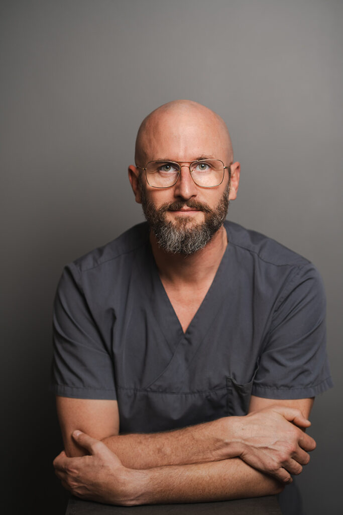
Felix Gussmann
Specialist in dermatology
- Aesthetic dermatology
- Outpatient operations
- Dermatooncology (skin cancer)
- Excessive sweating (hyperhidrosis)
- Specialist training at the University Clinic for Dermatology, Venereology and Allergology (under Prof. Dr. med. W. Sterry) at the Charité in Berlin
- Senior physician and deputy head of dermatosurgery at the dermatological clinic, Charité
- Head of the HIV consultation hour of the skin clinic, Charité

Lena Eydam
Specialist in dermatology
- Outpatient operations
- Dermatooncology (skin cancer)
- Aesthetic dermatology
- Phlebology
- – Licence to practise medicine – Charité Universitätsmedizin Berlin
–
Assistant doctor – Sankt Gertrauden-Krankenhaus GmbH – Berlin – Visceral surgery
–
Assistant doctor – Havelklinik Berlin – Dermatosurgery / Phlebology
– Recognised as a specialist in dermatology and venereology – Berlin Medical Association
As a long-standing member of various professional societies, such as the Network – Globalhealth, the DGDC (German Society for Dermatosurgery), the ISAC (International Society for Aesthetic Competence, formerly DEGAuF – German Society for Aesthetic Medicine and Training), the DDG (German Dermatological Society), the BVDD (Professional Association of German Dermatologists) and the GAERID (Society for Aesthetic and Reconstructive Intimate Surgery Germany e.V.). ), the DDG (Deutsche Dermatologische Gesellschaft), the BVDD (Berufsverband der Deutschen Dermatologen) and the GAERID (Gesellschaft für aesthetische und rekonstruktive Intimchirurgie Deutschland e.V.), we attend national and international congresses and regularly take part in further training courses in order to provide you with modern medicine and personalised, attentive treatment at all times.
Fotona 4D Laser
Dear patients, We can offer you another innovation in the field of aesthetic laser therapy. Don’t wait any longer to improve your skin. Contact us today to make an appointment for your Fotona 4D laser treatment and take the first step towards a younger, more radiant complexion. Would you like to improve your skin’s appearance … Continue reading "Fotona 4D Laser"
Read MoreContact & Directions
Our consultation hours
| Monday | 9:00 – 13:0015:00 – 18:00 |
|---|---|
| Tuesday | 9:00 – 13:0015:00 – 18:00 |
| Wednesday | 9:00 – 13:00 |
| Thursday | 9:00 – 13:0015:00 – 18:00 |
| Friday | 9:00 – 13:00 |
and by appointment
How to reach us
Dermatologische Praxis B18
Gussmann + Eydam
Badensche Strasse 18
10715 Berlin-Wilmersdorf
Phone: 030 814 869 30
Fax: 030 814 869 31
E-Mail: info@dermatologen-b18.de
Transport connection
U-Bahn: U7, U9 to U-Station Berliner Straße
Approach: Gesundheitszentrum B18, Badensche Straße 18, Ecke Prinzregentenstraße, Berlin-Wilmersdorf
It goes without saying that our house is handicapped accessible.

Show map
To activate the embedded map, please click on the link. By activating the map, data will be transmitted to the respective provider. Further information can be found in our privacy policy.
Imprint Privacy policy Cookie-Options
© 2025 by Dermatologists B-18 - Gussmann + Eydam. All rights reserved.
Made by wolter & works
